|
Carrots may be the food best known for helping your eyes. But other foods and their nutrients may be more important for keeping your eyesight keen as you age.
Vitamins C and E, zinc, lutein, zeaxanthin, and omega-3 fatty acids all play a role in eye health. They can help prevent cataracts. They may also fight the most-likely cause of vision loss when you're older: age-related macular degeneration (AMD). Here are some powerhouse foods for healthy eyes to try.
0 Comments
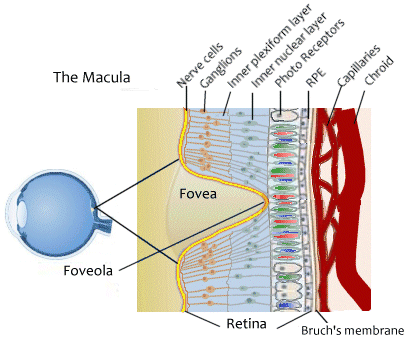
 Age-related macular degeneration (AMD or ARMD) is the most common cause of vision loss in those aged over 50. It causes a gradual loss of central (but not peripheral) vision. Central vision is required for detailed work and for tasks like reading and driving. The disease does not lead to complete blindness. Visual loss can occur within months, or over many years, depending on the type and severity of AMD. There are two main types of AMD - 'wet' and 'dry'. 'Wet' AMD is most severe but more treatable. Visual loss caused by AMD cannot normally be reversed. New medicines are an exciting development for wet AMD as they may halt or delay the progression of visual loss. Understanding the back of the eye · The retina is made up of two main layers. There is a layer of 'seeing cells' called rods and cones. These cells react to light and send electrical signals down tiny nerve fibres (which collect into the optic nerve) to the brain. The outer layer - the retinal pigment epithelium (RPE) - is a layer of cells behind the rods and cones. The RPE is an insulating layer between the retina and the choroid. These cells help to nourish and support the rods and cones. They pass nutrients from the blood vessels in the choroid to the rods and cones. They also take waste materials from the rods and cones to the blood vessels in the choroid. The RPE can be thought of as a filter, determining what substances reach the retina. Many components of blood are harmful to the retina and are kept away from it by a normally functioning RPE. The rods and cones are responsible for vision in different conditions. There are many more rods than cones, and rods are smaller cells than cones: · The cone cells ('cones') help us to see in the daylight, providing the basis for colour vision. · The rod cells ('rods') help us to see in the dark - 'night vision'. · The macula is a small but vital area of the retina at the back of your eye. It is about 5 mm in diameter. The macula is the part of the retina that is the most densely packed with rods and cones. The macula is essential for central vision. In the middle of the macula is an area called the fovea, which only contains cones. · The choroid is a layer of tissue behind the retina which contains many tiny blood vessels. These help to take oxygen and nutrients to the retina. · Bruch's membrane is a thin membrane which helps to form a barrier between the choroid and the delicate retina. · The sclera is the outer thick white layer of the eye. When you look at an object, light from the object passes through the cornea, then the lens, and then hits the retina at the back of the eye. The light from the object focuses on the macula. You need a healthy macula for detailed central vision. What is age-related macular degeneration? AMD is a condition that occurs when cells in the macula degenerate. This occurs with partial breakdown of the RPE and the cells become damaged and die. Damage to the macula affects your central vision which is needed for reading, writing, driving, recognising people's faces and doing other fine tasks. The rest of the retina is used for peripheral vision - the 'side' vision which is not focused. Therefore, without a macula you can still see enough to get about, be aware of objects and people, and be independent. However, the loss of central vision will severely affect normal sight. There are two types - 'dry' and 'wet' AMD - described below. Who gets age-related macular degeneration? AMD is the most common form of macular degeneration and develops in older people. There are other rare types of macular degeneration which occur in younger people. AMD can affect anyone. It is the most common cause of severe sight problems (visual impairment) in the UK, and indeed in the developed world. It becomes more common with increasing age, as the name suggests. It is rare under the age of 60. If you develop wet AMD (see below) in one eye the risk of developing wet AMD in the second eye is about 1 in 4. About 5 in 100 people aged over 65 and about 1 in 8 people aged over 80 have AMD severe enough to cause serious visual loss. About twice as many women over the age of 75 have AMD compared with men of the same age. The two types of age-related macular degeneration Dry AMD This is the most common form and occurs in 9 in 10 cases. In this type the cells in the RPE of the macula gradually become thin (they 'atrophy') and degenerate. This layer of cells is crucial for the function of the rods and cones which then also degenerate and die. Typically, dry AMD is a very gradual process as the number of cells affected increases. It usually takes several years for vision to become seriously affected. Many people with dry AMD do not totally lose their reading vision. Wet AMD Wet AMD may also be called neovascular or exudative AMD. It occurs in about 1 in 10 cases. However, it is likely to cause severe visual loss over quite a short time - sometimes just months. Very occasionally, if there is a bleed (haemorrhage) from a new blood vessel, this visual loss can occur suddenly, within hours or days. In wet AMD, in addition to the retinal pigment cells degenerating, new tiny blood vessels grow from the tiny blood vessels in the choroid. This is called choroidal neovascularisation. The new vessels break through Bruch's membrane and into the macular part of the retina. These vessels are not normal. They are fragile and tend to leak blood and fluid. This can damage the rods and cones, and cause scarring in the macula, causing further vision loss. Both wet and dry AMD are further classified according to severity. Early, intermediate or advanced types refer to the degree of damage to the macula. 6 in 10 cases of intermediate/advanced AMD are due to wet AMD. What causes age-related macular degeneration? In people with AMD the cells of the RPE do not work so well with advancing age. They gradually fail to take enough nutrients to the rods and cones, and do not clear waste materials and byproducts made by the rods and cones either. As a result, tiny abnormal deposits called drusen develop under the retina. In time, the retinal pigment cells and their nearby rods and cones degenerate, stop working and die. This is the dry type of AMD. In other cases, something also triggers new blood vessels to develop from the choroid to cause the wet form of AMD. The trigger is not known. It may be that some waste products which are not cleared from the RPE may stimulate new blood vessels to grow in an attempt to clear the waste. The exact reason why cells of the RPE stop working properly in people with AMD is not known. Certain risk factors increase the risk of developing AMD. These include: · Smoking tobacco. · Possibly, high blood pressure (inconclusive evidence). · A family history of AMD. (AMD is not a straightforward hereditary condition. However, your risk of developing AMD is increased if it occurs in other family members.) · Sunlight. This has yet to be proven, but laboratory studies suggest that the retina is damaged by sunlight rays (UVA and UVB rays). AMD seems to be more common in people from white (Caucasian) racial backgrounds than from other racial groups. What are the symptoms of age-related macular degeneration? · The main early symptom is blurring of central vision despite using your usual glasses. In the early stages of the condition you may notice that: · You need brighter light to read by. · Words in a book or newspaper may become blurred. · Colours appear less bright. · You have difficulty recognising faces. · One specific early symptom to be aware of is visual distortion. Typically, straight lines appear wavy or crooked. For example, the lines on a piece of graph paper, or the lines between tiles in a bathroom, or the border of any other straight object, etc. · A 'blind spot' then develops in the middle of your visual field. This tends to become larger over time as more and more rods and cones degenerate in the macula. · Visual hallucinations are common in people with severe visual loss of any cause. Visual hallucinations (also called Charles Bonnet syndrome) can occur if you have severe AMD. People see different images, from simple patterns to more detailed pictures. The experience can be upsetting but is less frightening if you are aware that it can happen in AMD. Importantly, it does not mean you are developing a serious mental illness. If you do develop visual hallucinations they typically improve by 18 months but in some people they last for years. AMD is painless. Symptoms of dry AMD tend to take 5-10 years to become severe. However, severe visual loss due to wet AMD can develop more quickly. Always see a doctor or optometrist promptly if you develop visual loss or visual distortion. This is not only the case if you are worried about AMD. Other sight-threatening conditions can occur suddenly with visual loss, such as a detached retina. Peripheral vision is not affected with AMD and so it does not cause total blindness. Note: if the vision of one eye only is affected, you may not notice any symptoms, as the other good eye often compensates. When both eyes are affected you are more likely to notice symptoms. Older people should have regular eye checks to check each eye separately for early AMD (and to check for other eye conditions such as glaucoma). How is age-related macular degeneration diagnosed? If you develop symptoms suggestive of AMD, we will examine the back of your eye with a slit lamp microscope. This is a magnifying piece of equipment which the optometrist uses to examine your retinae through what look like binoculars. Another test called ocular coherence tomography is also very useful. This is a non-invasive test that uses special light rays to scan the retina. It can give very detailed '3D' information about the macula, and can show if the macula is thickened or abnormal. This test is useful when there is doubt about whether AMD is the wet or dry form. It is also a useful test to assess and monitor the results of any treatment. Practical help When your vision becomes poor, it is common to be referred (by your optometrist) to a low vision clinic. Staff at the clinic provide practical help and advice on how to cope with poor and/or deteriorating vision. Help may include: · Magnifying lenses, large print books, and bright lamps which may assist reading. · Gadgets such as talking watches and kitchen aids which can help when vision is limited. · Being registered as partially sighted or blind. Your consultant ophthalmologist can complete a 'Certificate of Visual Impairment'. You may then be entitled to certain benefits. What else can I do? · If you smoke, try to stop. If you are smoker, there are numerous health benefits to quitting. Smoking is a risk factor for many illnesses, including AMD. The NHS can provide help, support and medicines to assist stopping smoking. · Eat a healthy balanced diet to try to make sure you get plenty of the types of vitamins that may help in AMD. · Stay safe with regards to driving. If you are registered with sight impairment you should not drive and should notify the Driver and Vehicle Licensing Agency (DVLA). The DVLA provides detailed guidance on fitness to drive and minimum standards with regard to sight. This includes being able to read, wearing your normal glasses, a vehicle number plate at a distance of 20 metres. · Consider regular sight tests as you get older. You should visit an optometrist every two years, even if there is no change in your vision. An eye test can often pick up the first signs of an eye condition before you notice any change in your vision. Your optometrist can advise you how often you need to have an eye check-up, depending on your general health, age, family history and other medical conditions. Early detection of problems often allows more effective treatment. To book an appointment call us on 01268 544646. This Christmas when you are planning your meals, or enjoying them, think about how they are affecting your eyes. Don't leave it until New Years Day to start eating healthy because you can be naughty and nice!
We frequently take our eyes for granted, but these are highly specialised organs that require careful maintenance to operate at their optimal capacity. While eye tests and vision correction products play key roles In this process, the foods we eat can also be greatly beneficial. Studies around the world have emphasised that a healthy lifestyle combined with healthy eating can reduce the prevalence of cataracts, while carbohydrate-high, vitamin-low diets directly increase this risk. Similarly, a carefully balanced diet helps to counteract age-related macular degeneration, or AMD. This is the leading cause of registered blindness in the western world, but can be halted and even partly reversed through prompt diagnosis and positive lifestyle choices. Research has established that obesity can double the risk of developing some common causes of blindness, including AMD. Although our retinas naturally weaken over time excess body weight can dramatically speed up the onset of AMD, giving us yet another reason to consider what we eat and how it might affect our bodies. For many years, the focus on diet and its impact on our vision have concentrated on vitamins A, C and E. Numerous scientific studies and clinical trials have shown that these three ingredients help to maintain healthy cells and tissues in our eyes, even assisting with our tear functions and reducing the symptoms of dry eyes. Should your diet not lend itself to a regular intake of fresh produce, nutritional supplements can top up many missing vitamins and minerals although use of these supplements should ideally be approved by your GP. So when you have the turkey add lots of vegetables to the mix and start protecting your eyes now! |
Author:
|
|
Templeman Opticians -Laindon,
Danacre, Laindon, Essex. SS15 5PS |
© Copyright Templeman Opticians Laindon 2023
|


 RSS Feed

